Case of the month
The Case of the Month aims to share interesting and educational genito-urinary pathology cases with the ISUP membership. All ISUP members are cordially invited to submit cases to the case manager (cotm@isupweb.org) using the available template (word document). Your submission is highly appreciated!
May 2017
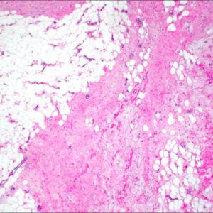
A 54 year old male with past medical history of hydrocele, presents with onset of bilateral scrotal masses and buried penis 6 months status post uncomplicated left inguinal herniorrhaphy. Sonographic imaging demonstrated skin thinking with underlying amorphous subcutaneous mass involving the scrotum, left > right, with compression of and atrophy of the left testis. Patient underwent resection of bilateral scrotal masses, which measured 21×16.5×4.5cm and 14x9x3.5cm.
April 2017
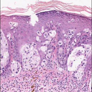
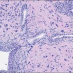
81 year-old man presenting with an isolated leukoplakia-like penile (preputial) lesion of unknown duration.
A 0.3 cm incisional biopsy was performed.

March 2017
A 73-year- old male with LUTS and s-PSA 6.6. A 10-core prostate biopsy was taken. In addition to a conventional prostate cancer GS 7 (neg for p63, pos for AMACR), a glandular proliferation is seen in 4 cores.
February 2017
A 42-year-old male with right ureteric stricture
4-year history of recurrent right ureteric calculi, status post ureteroscopic lithotripsy (USRL) twice
Cystoscopy: Stenosis of right ureteric orifice, which was unable to pass guide wire
Treatment: Ureteric reimplantation with lysis of adhesion was performed; ureteric wall was sent to pathology
Gross findings: Thickened ureteric wall lined by smooth and flat mucosa

January 2017
44-year-old man with right renal mass, retroperitoneal and mediastinal lymphadenopathy
An adrenal-sparing left radical nephrectomy was performed
Patient underwent several cycles of chemotherapy and biological treatment
Patient is alive with residual disease (retroperitoneal lymph nodes) 9 years after nephrectomy
December 2016
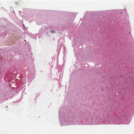
71 year-old male with a 3.4 cm, mid-pole, left renal mass
An adrenal-sparing left radical nephrectomy was performed
Gross Description
Radical nephrectomy specimen
Single 3.4 cm yellow-brown, focally hemorrhagic mass in the mid-pole aspect of the kidney
Tumour grossly confined to the kidney – abutting the middle renal calyx and bulging into the renal sinus
No involvement of the renal vein or its segmental branches

November 2016
A 68 year-old male with HIV and clinical benign prostatic hyperplasia underwent TURP.
In one of the TURP chips a glandular proliferation is seen streaming between benign glands.

October 2016
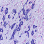
A 53 year-old female with an incidentally found 2.9 cm right renal mass on abdominal ultrasound.
Percutaneous needle biopsy was performed
limited lesion sampling (1-2 mm of tumor in a 15 mm core)
bland ovoid and spindle cells with no overt malignant features
positive for CD34 and SMA; negative for epithelial markers
reported as “mesenchymal neoplasm – definitive classification not possible on biopsy”
Patient chose surgical management over active surveillance with serial imaging
A right partial nephrectomy was performed
JULY 2016
– 55 year old male with long term use of exogenous testosterone.
– PSA- 6.9 ng/ml
– Digital rectal exam – large, firm prostate gland
– U/S imaging – anterior nodule, possible extraprostatic disease
– TRUS guided biopsies – 12 core set, anterior nodule not targeted for biopsy

June 2016
64 year old male
5 year history of intermittent gross hematuria
Social history: Non-smoker, metal plater with exposure to trichloroethylene 15-20 years ago
No history of previous bladder lesions
3.5 cm bladder neck / trigone tumor
Cystoscopic examination: Papillary tumor with a narrow stalk, suspicious for urothelial carcinoma
Treatment: Transurethral resection of tumor